Electrocardiogram Tunisia
Electrocardiogram or ECG is a recording of heart activity. This test can diagnose an arrhythmia or coronary disease, such as heart disease. It can also be used to understand the cause of certain conditions, such as chest or heart pain, that may be caused by the heart.
The doctor will place ten electrodes on your skin: six electrodes on your chest and one at each end of your limbs. The traces of the heart's impulse have appeared on the paper. The examination by the cardiologist or general practitioner at the doctor's office takes 10 minutes.

The Holter simply records the ECG continuously for 24 or 48 hours. When there are no pit symptoms during the resting ECG, a prescription may be prescribed. It is used to detect intermittent arrhythmia, to detect heart rhythm abnormalities, especially if there are symptoms such as palpitations or syncopation (faintness with loss of consciousness), and to adjust medication treatment for known heart rhythm disorders.
The electrodes, which enable the electrical recording of cardiac activity, are attached to the trunk with adhesive tape.
After analyzing the results, the doctor can make a diagnosis of rhythm disorders.
Note: This examination is usually performed in addition to an electrocardiogram.
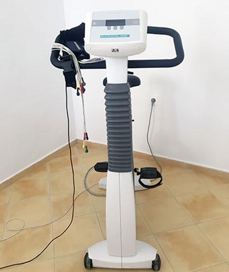
The stress test refers to the recording of the electrocardiogram during the performance of a calibrated physical effort under medical supervision. In fact, heart problems sometimes occur only during physical activity.
An abnormally calm heart, arrhythmia and heart disease (such as angina pectoris). It is also useful to measure the athletic ability of people over 40 years of age who wish to resume physical activity.
The doctor puts electrodes on your chest and then you ride your bike. During the recording process, under the supervision of the medical team, the intensity of the work gradually increases.
This ultrasound technique for detecting the heart allows real-time observation of heart muscle contraction and valve movement.
This inspection is usually combined with Doppler. This allows you to see the blood flow in the chambers of the heart and the large blood vessels (artery, vena cava, pulmonary arteries and veins).
Echocardiography Tunisia can detect certain abnormalities, such as valve infections or other heart diseases. It can also detect the presence of blood clots or detect heart infections.
After applying a lot of gel, the echographer will move the probe around your chest. The exam takes about 20 minutes.
Ultrasound is a non-invasive medical technology that can probe the human body through a process similar to the use of a sonarultrasound.
The Doppler effect can measure the movement of red blood cells in the blood vessel itself, so it can be used to visualize blood flow in arteries and veins.
Doppler ultrasound combines the ultrasound and Doppler effects. The examination usually lasts 15 minutes and the operator must be very concentrated and vigilant.
